ACTG2
Protein-coding gene in the species Homo sapiens
| ACTG2 | |||||||||||||||||||||||||||||||||||||||||||||||||||
|---|---|---|---|---|---|---|---|---|---|---|---|---|---|---|---|---|---|---|---|---|---|---|---|---|---|---|---|---|---|---|---|---|---|---|---|---|---|---|---|---|---|---|---|---|---|---|---|---|---|---|---|
 | |||||||||||||||||||||||||||||||||||||||||||||||||||
| Identifiers | |||||||||||||||||||||||||||||||||||||||||||||||||||
| Aliases | ACTG2, ACT, ACTA3, ACTE, ACTL3, ACTSG, VSCM, actin, gamma 2, smooth muscle, enteric, actin gamma 2, smooth muscle, VSCM1, MMIHS5 | ||||||||||||||||||||||||||||||||||||||||||||||||||
| External IDs | OMIM: 102545; MGI: 104589; HomoloGene: 123845; GeneCards: ACTG2; OMA:ACTG2 - orthologs | ||||||||||||||||||||||||||||||||||||||||||||||||||
| |||||||||||||||||||||||||||||||||||||||||||||||||||
| |||||||||||||||||||||||||||||||||||||||||||||||||||
| |||||||||||||||||||||||||||||||||||||||||||||||||||
| |||||||||||||||||||||||||||||||||||||||||||||||||||
| |||||||||||||||||||||||||||||||||||||||||||||||||||
| Wikidata | |||||||||||||||||||||||||||||||||||||||||||||||||||
| |||||||||||||||||||||||||||||||||||||||||||||||||||


Actin, gamma-enteric smooth muscle is a protein that in humans is encoded by the ACTG2 gene.[5][6][7]
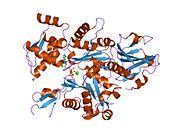
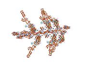
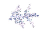
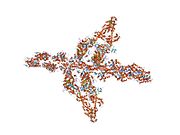
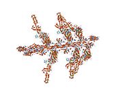
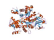
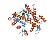
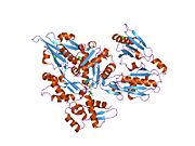
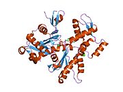
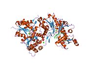
Actins are highly conserved proteins that are involved in various types of cell motility, and maintenance of the cytoskeleton. In vertebrates, three main groups of actin isoforms, alpha, beta and gamma have been identified. The alpha actins are found in muscle tissues and are a major constituent of the contractile apparatus. The beta and gamma actins co-exist in most cell types as components of the cytoskeleton, and as mediators of internal cell motility. Actin, gamma 2, encoded by this gene, is a smooth muscle actin found in enteric tissues.[7]
Interactions
ACTG2 has been shown to interact with Emerin.[8]
References
- ^ a b c GRCh38: Ensembl release 89: ENSG00000163017 – Ensembl, May 2017
- ^ a b c GRCm38: Ensembl release 89: ENSMUSG00000059430 – Ensembl, May 2017
- ^ "Human PubMed Reference:". National Center for Biotechnology Information, U.S. National Library of Medicine.
- ^ "Mouse PubMed Reference:". National Center for Biotechnology Information, U.S. National Library of Medicine.
- ^ Miwa T, Manabe Y, Kurokawa K, Kamada S, Kanda N, Bruns G, Ueyama H, Kakunaga T (July 1991). "Structure, chromosome location, and expression of the human smooth muscle (enteric type) gamma-actin gene: evolution of six human actin genes". Mol Cell Biol. 11 (6): 3296–306. doi:10.1128/mcb.11.6.3296. PMC 360182. PMID 1710027.
- ^ Ueyama H (May 1991). "A HindIII DNA polymorphism in the human enteric type smooth muscle actin gene (ACTSG)". Nucleic Acids Res. 19 (2): 411. doi:10.1093/nar/19.2.411. PMC 333620. PMID 1673027.
- ^ a b "Entrez Gene: ACTG2 actin, gamma 2, smooth muscle, enteric".
- ^ Lattanzi G, Cenni Vittoria, Marmiroli Sandra, Capanni Cristina, Mattioli Elisabetta, Merlini Luciano, Squarzoni Stefano, Maraldi Nadir Mario (April 2003). "Association of emerin with nuclear and cytoplasmic actin is regulated in differentiating myoblasts". Biochem. Biophys. Res. Commun. 303 (3). United States: 764–70. doi:10.1016/S0006-291X(03)00415-7. ISSN 0006-291X. PMID 12670476.
External links
- Human ACTG2 genome location and ACTG2 gene details page in the UCSC Genome Browser.
Further reading
- Snásel J, Pichová I (1997). "The cleavage of host cell proteins by HIV-1 protease". Folia Biol. (Praha). 42 (5): 227–30. doi:10.1007/BF02818986. PMID 8997639. S2CID 7617882.
- Adams LD, Tomasselli AG, Robbins P, et al. (1992). "HIV-1 protease cleaves actin during acute infection of human T-lymphocytes". AIDS Res. Hum. Retroviruses. 8 (2): 291–5. doi:10.1089/aid.1992.8.291. PMID 1540415.
- Tomasselli AG, Hui JO, Adams L, et al. (1991). "Actin, troponin C, Alzheimer amyloid precursor protein and pro-interleukin 1 beta as substrates of the protease from human immunodeficiency virus". J. Biol. Chem. 266 (22): 14548–53. doi:10.1016/S0021-9258(18)98721-1. PMID 1907279.
- Shoeman RL, Kesselmier C, Mothes E, et al. (1991). "Non-viral cellular substrates for human immunodeficiency virus type 1 protease". FEBS Lett. 278 (2): 199–203. Bibcode:1991FEBSL.278..199S. doi:10.1016/0014-5793(91)80116-K. PMID 1991513. S2CID 37002682.
- Miwa T, Kamada S (1990). "The nucleotide sequence of a human smooth muscle (enteric type) gamma-actin cDNA". Nucleic Acids Res. 18 (14): 4263. doi:10.1093/nar/18.14.4263. PMC 331204. PMID 2377475.
- Ueyama H, Inazawa J, Nishino H, et al. (1995). "Chromosomal mapping of the human smooth muscle actin gene (enteric type, ACTA3) to 2p13.1 and molecular nature of the hindIII polymorphism". Genomics. 25 (3): 720–3. doi:10.1016/0888-7543(95)80016-F. PMID 7759108.
- Szucsik JC, Lessard JL (1996). "Cloning and sequence analysis of the mouse smooth muscle gamma-enteric actin gene". Genomics. 28 (2): 154–62. doi:10.1006/geno.1995.1126. PMID 8530021.
- Bukrinskaya A, Brichacek B, Mann A, Stevenson M (1999). "Establishment of a Functional Human Immunodeficiency Virus Type 1 (HIV-1) Reverse Transcription Complex Involves the Cytoskeleton". J. Exp. Med. 188 (11): 2113–25. doi:10.1084/jem.188.11.2113. PMC 2212381. PMID 9841925.
- Zhang H, Wang L, Kao S, et al. (1999). "Functional interaction between the cytoplasmic leucine-zipper domain of HIV-1 gp41 and p115-RhoGEF". Curr. Biol. 9 (21): 1271–4. Bibcode:1999CBio....9.1271Z. doi:10.1016/S0960-9822(99)80511-9. PMC 4513661. PMID 10556093.
- Kohnen G, Campbell S, Jeffers MD, Cameron IT (2000). "Spatially regulated differentiation of endometrial vascular smooth muscle cells". Hum. Reprod. 15 (2): 284–92. doi:10.1093/humrep/15.2.284. PMID 10655297.
- Filmore RA, Dean DA, Zimmer WE (2003). "The smooth muscle gamma-actin gene is androgen responsive in prostate epithelia". Gene Expr. 10 (5–6): 201–11. doi:10.3727/000000002783992424 (inactive 31 January 2024). PMC 5977519. PMID 12450213.
{{cite journal}}: CS1 maint: DOI inactive as of January 2024 (link) - Strausberg RL, Feingold EA, Grouse LH, et al. (2003). "Generation and initial analysis of more than 15,000 full-length human and mouse cDNA sequences". Proc. Natl. Acad. Sci. U.S.A. 99 (26): 16899–903. Bibcode:2002PNAS...9916899M. doi:10.1073/pnas.242603899. PMC 139241. PMID 12477932.
- Cheng C, Sharp PA (2003). "RNA Polymerase II Accumulation in the Promoter-Proximal Region of the Dihydrofolate Reductase and γ-Actin Genes". Mol. Cell. Biol. 23 (6): 1961–7. doi:10.1128/MCB.23.6.1961-1967.2003. PMC 149466. PMID 12612070.
- Wu RF, Gu Y, Xu YC, et al. (2004). "Human Immunodeficiency Virus Type 1 Tat Regulates Endothelial Cell Actin Cytoskeletal Dynamics through PAK1 Activation and Oxidant Production". J. Virol. 78 (2): 779–89. doi:10.1128/JVI.78.2.779-789.2004. PMC 368750. PMID 14694110.
- Ota T, Suzuki Y, Nishikawa T, et al. (2004). "Complete sequencing and characterization of 21,243 full-length human cDNAs". Nat. Genet. 36 (1): 40–5. doi:10.1038/ng1285. PMID 14702039.
- Gerhard DS, Wagner L, Feingold EA, et al. (2004). "The Status, Quality, and Expansion of the NIH Full-Length cDNA Project: The Mammalian Gene Collection (MGC)". Genome Res. 14 (10B): 2121–7. doi:10.1101/gr.2596504. PMC 528928. PMID 15489334.
- Hillier LW, Graves TA, Fulton RS, et al. (2005). "Generation and annotation of the DNA sequences of human chromosomes 2 and 4". Nature. 434 (7034): 724–31. Bibcode:2005Natur.434..724H. doi:10.1038/nature03466. PMID 15815621.
- v
- t
- e
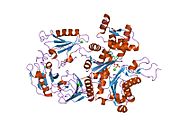
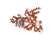
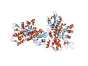
PDB gallery
-
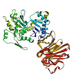
 1atn: Atomic structure of the actin:DNASE I complex
 -
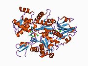
 1c0f: CRYSTAL STRUCTURE OF DICTYOSTELIUM CAATP-ACTIN IN COMPLEX WITH GELSOLIN SEGMENT 1
 -
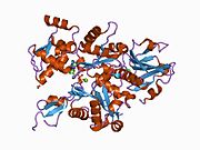
 1c0g: CRYSTAL STRUCTURE OF 1:1 COMPLEX BETWEEN GELSOLIN SEGMENT 1 AND A DICTYOSTELIUM/TETRAHYMENA CHIMERA ACTIN (MUTANT 228: Q228K/T229A/A230Y/E360H)
 -
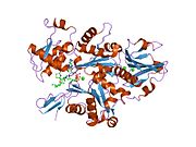
 1d4x: Crystal Structure of Caenorhabditis Elegans Mg-ATP Actin Complexed with Human Gelsolin Segment 1 at 1.75 A resolution.
 -
 1dej: CRYSTAL STRUCTURE OF A DICTYOSTELIUM/TETRAHYMENA CHIMERA ACTIN (MUTANT 646: Q228K/T229A/A230Y/A231K/S232E/E360H) IN COMPLEX WITH HUMAN GELSOLIN SEGMENT 1
 -
 1eqy: COMPLEX BETWEEN RABBIT MUSCLE ALPHA-ACTIN: HUMAN GELSOLIN DOMAIN 1
 -
 1esv: COMPLEX BETWEEN LATRUNCULIN A:RABBIT MUSCLE ALPHA ACTIN:HUMAN GELSOLIN DOMAIN 1
 -
 1h1v: GELSOLIN G4-G6/ACTIN COMPLEX
 -
 1hlu: STRUCTURE OF BOVINE BETA-ACTIN-PROFILIN COMPLEX WITH ACTIN BOUND ATP PHOSPHATES SOLVENT ACCESSIBLE
 -
 1ijj: THE X-RAY CRYSTAL STRUCTURE OF THE COMPLEX BETWEEN RABBIT SKELETAL MUSCLE ACTIN AND LATRUNCULIN A AT 2.85 A RESOLUTION
 -
 1j6z: UNCOMPLEXED ACTIN
 -
 1kxp: CRYSTAL STRUCTURE OF HUMAN VITAMIN D-BINDING PROTEIN IN COMPLEX WITH SKELETAL ACTIN
 -
 1lcu: Polylysine Induces an Antiparallel Actin Dimer that Nucleates Filament Assembly: Crystal Structure at 3.5 A Resolution
 -
 1lot: CRYSTAL STRUCTURE OF THE COMPLEX OF ACTIN WITH VITAMIN D-BINDING PROTEIN
 -
 1m8q: Molecular Models of Averaged Rigor Crossbridges from Tomograms of Insect Flight Muscle
 -
 1ma9: Crystal structure of the complex of human vitamin D binding protein and rabbit muscle actin
 -
 1mdu: Crystal structure of the chicken actin trimer complexed with human gelsolin segment 1 (GS-1)
 -
 1mvw: MOLECULAR MODELS OF AVERAGED RIGOR CROSSBRIDGES FROM TOMOGRAMS OF INSECT FLIGHT MUSCLE
 -
 1nlv: Crystal Structure Of Dictyostelium Discoideum Actin Complexed With Ca ATP And Human Gelsolin Segment 1
 -
 1nm1: Crystal Structure of D. Dicsoideum Actin Complexed With Gelsolin Segment 1 and Mg ATP at 1.8 A Resolution
 -
 1nmd: Crystal Structure of D. Discoideum Actin-Gelsolin Segment 1 Complex Crystallized In Presence Of Lithium ATP
 -
 1nwk: CRYSTAL STRUCTURE OF MONOMERIC ACTIN IN THE ATP STATE
 -
 1o18: MOLECULAR MODELS OF AVERAGED RIGOR CROSSBRIDGES FROM TOMOGRAMS OF INSECT FLIGHT MUSCLE
 -
 1o19: MOLECULAR MODELS OF AVERAGED RIGOR CROSSBRIDGES FROM TOMOGRAMS OF INSECT FLIGHT MUSCLE
 -
 1o1a: MOLECULAR MODELS OF AVERAGED RIGOR CROSSBRIDGES FROM TOMOGRAMS OF INSECT FLIGHT MUSCLE
 -
 1o1b: MOLECULAR MODELS OF AVERAGED RIGOR CROSSBRIDGES FROM TOMOGRAMS OF INSECT FLIGHT MUSCLE
 -
 1o1c: MOLECULAR MODELS OF AVERAGED RIGOR CROSSBRIDGES FROM TOMOGRAMS OF INSECT FLIGHT MUSCLE
 -
 1o1d: MOLECULAR MODELS OF AVERAGED RIGOR CROSSBRIDGES FROM TOMOGRAMS OF INSECT FLIGHT MUSCLE
 -
 1o1e: MOLECULAR MODELS OF AVERAGED RIGOR CROSSBRIDGES FROM TOMOGRAMS OF INSECT FLIGHT MUSCLE
 -
 1o1f: MOLECULAR MODELS OF AVERAGED RIGOR CROSSBRIDGES FROM TOMOGRAMS OF INSECT FLIGHT MUSCLE
 -
 1o1g: MOLECULAR MODELS OF AVERAGED RIGOR CROSSBRIDGES FROM TOMOGRAMS OF INSECT FLIGHT MUSCLE
 -
 1p8z: Complex Between Rabbit Muscle alpha-Actin: Human Gelsolin Residues Val26-Glu156
 -
 1qz5: Structure of rabbit actin in complex with kabiramide C
 -
 1qz6: Structure of rabbit actin in complex with jaspisamide A
 -
 1rdw: Actin Crystal Dynamics: Structural Implications for F-actin Nucleation, Polymerization and Branching Mediated by the Anti-parallel Dimer
 -
 1rfq: Actin Crystal Dynamics: Structural Implications for F-actin Nucleation, Polymerization and Branching Mediated by the Anti-parallel Dimer
 -
 1rgi: Crystal structure of gelsolin domains G1-G3 bound to actin
 -
 1s22: Absolute Stereochemistry of Ulapualide A
 -
 1sqk: CRYSTAL STRUCTURE OF CIBOULOT IN COMPLEX WITH SKELETAL ACTIN
 -
 1t44: Structural basis of actin sequestration by thymosin-B4: Implications for arp2/3 activation
 -
 1wua: The structure of Aplyronine A-actin complex
 -
 1y64: Bni1p Formin Homology 2 Domain complexed with ATP-actin
 -
 1yxq: Crystal structure of actin in complex with swinholide A
 -
 2a3z: Ternary complex of the WH2 domain of WASP with Actin-DNAse I
 -
 2a40: Ternary complex of the WH2 domain of WAVE with Actin-DNAse I
 -
 2a41: Ternary complex of the WH2 Domain of WIP with Actin-DNAse I
 -
 2a42: Actin-DNAse I Complex
 -
 2a5x: Crystal Structure of a Cross-linked Actin Dimer
 -
 2asm: Structure of Rabbit Actin In Complex With Reidispongiolide A
 -
 2aso: Structure of Rabbit Actin In Complex With Sphinxolide B
 -
 2asp: Structure of Rabbit Actin In Complex With Reidispongiolide C
 -
 2btf: THE STRUCTURE OF CRYSTALLINE PROFILIN-BETA-ACTIN
 -
 2d1k: Ternary complex of the WH2 domain of mim with actin-dnase I
 -
 2ff3: Crystal structure of Gelsolin domain 1:N-wasp V2 motif hybrid in complex with actin
 -
 2ff6: Crystal structure of Gelsolin domain 1:ciboulot domain 2 hybrid in complex with actin
 -
 2fxu: X-ray Structure of Bistramide A- Actin Complex at 1.35 A resolution.
 -
 2gwj: SpvB ADP-ribosylated actin: hexagonal crystal form
 -
 2gwk: SpvB ADP-ribosylated actin: orthorhombic crystal form
 -
 2hf3: Crystal structure of monomeric Actin in the ADP bound state
 -
 2hf4: Crystal structure of Monomeric Actin in its ATP-bound state
 -
 2hmp: Uncomplexed actin cleaved with protease ECP32
 -
 2oan: Structure of oxidized beta-actin
 -
 2q1n: Actin Dimer Cross-linked Between Residues 41 and 374
 -
 2q31: Actin Dimer Cross-linked Between Residues 41 and 374 and proteolytically cleaved by subtilisin between residues 47 and 48.
 -
 2q36: Actin Dimer Cross-linked between Residues 191 and 374 and complexed with Kabiramide C


 | This article on a gene on human chromosome 2 is a stub. You can help Wikipedia by expanding it. |
- v
- t
- e


