Protein-coding gene in the species Homo sapiens
| B3GAT1 |
|---|
 |
| Available structures |
|---|
| PDB | Ortholog search: PDBe RCSB |
|---|
| List of PDB id codes |
|---|
1V82, 1V83, 1V84 |
|
|
| Identifiers |
|---|
| Aliases | B3GAT1, CD57, GLCATP, GLCUATP, HNK1, LEU7, NK-1, NK1, beta-1,3-glucuronyltransferase 1 |
|---|
| External IDs | OMIM: 151290 MGI: 1924148 HomoloGene: 49551 GeneCards: B3GAT1 |
|---|
| Gene location (Human) |
|---|
 | | Chr. | Chromosome 11 (human)[1] |
|---|
| | Band | 11q25 | Start | 134,378,504 bp[1] |
|---|
| End | 134,412,242 bp[1] |
|---|
|
| Gene location (Mouse) |
|---|
 | | Chr. | Chromosome 9 (mouse)[2] |
|---|
| | Band | 9|9 A4 | Start | 26,645,024 bp[2] |
|---|
| End | 26,674,397 bp[2] |
|---|
|
| RNA expression pattern |
|---|
| Bgee | | Human | Mouse (ortholog) |
|---|
| Top expressed in | - amygdala
- prefrontal cortex
- Brodmann area 9
- hippocampus proper
- putamen
- nucleus accumbens
- Region I of hippocampus proper
- caudate nucleus
- endothelial cell
- substantia nigra
|
| | Top expressed in | - habenula
- visual cortex
- piriform cortex
- superior frontal gyrus
- subiculum
- dentate gyrus
- superior colliculus
- Region I of hippocampus proper
- primary motor cortex
- medial dorsal nucleus
|
| | More reference expression data |
|
|---|
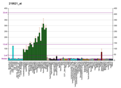
| BioGPS |  | | More reference expression data |
|
|---|
|
| Gene ontology |
|---|
| Molecular function | - transferase activity
- metal ion binding
- galactosylgalactosylxylosylprotein 3-beta-glucuronosyltransferase activity
- UDP-galactose:beta-N-acetylglucosamine beta-1,3-galactosyltransferase activity
| | Cellular component | - integral component of membrane
- Golgi apparatus
- membrane
- extracellular region
- endoplasmic reticulum
- endoplasmic reticulum membrane
- intracellular membrane-bounded organelle
- Golgi membrane
| | Biological process | - glycosaminoglycan metabolic process
- protein glycosylation
- chondroitin sulfate proteoglycan biosynthetic process
- chondroitin sulfate metabolic process
- carbohydrate metabolic process
- cellular response to hypoxia
| | Sources:Amigo / QuickGO |
|
| Orthologs |
|---|
| Species | Human | Mouse |
|---|
| Entrez | | |
|---|
| Ensembl | | |
|---|
| UniProt | | |
|---|
| RefSeq (mRNA) | |
|---|
NM_018644
NM_054025
NM_001367973 |
| |
|---|
| RefSeq (protein) | |
|---|
NP_061114
NP_473366
NP_001354902 |
| |
|---|
| Location (UCSC) | Chr 11: 134.38 – 134.41 Mb | Chr 9: 26.65 – 26.67 Mb |
|---|
| PubMed search | [3] | [4] |
|---|
|
| Wikidata |
| View/Edit Human | View/Edit Mouse |
|
3-beta-glucuronosyltransferase 1 (B3GAT1) is an enzyme that in humans is encoded by the B3GAT1 gene, whose enzymatic activity creates the CD57 epitope on other cell surface proteins.[5] In immunology, the CD57 antigen (CD stands for cluster of differentiation) is also known as HNK1 (human natural killer-1) or LEU7. It is expressed as a carbohydrate epitope that contains a sulfoglucuronyl residue in several adhesion molecules of the nervous system.[6]
Function
The protein encoded by this gene is a member of the glucuronyltransferase gene family. These enzymes exhibit strict acceptor specificity, recognizing nonreducing terminal sugars and their anomeric linkages. This gene product functions as the key enzyme in a glucuronyl transfer reaction during the biosynthesis of the carbohydrate epitope HNK-1 (human natural killer-1, also known as CD57 and LEU7). Alternate transcriptional splice variants have been characterized.[5]
Immunohistochemistry
In anatomical pathology, CD57 (immunostaining) is similar to CD56 for use in differentiating neuroendocrine tumors from others.[7] Using immunohistochemistry, CD57 molecule can be demonstrated in around 10 to 20% of lymphocytes, as well as in some epithelial, neural, and chromaffin cells. Among lymphocytes, CD57 positive cells are typically either T cells or NK cells, and are most commonly found within the germinal centres of lymph nodes, tonsils, and the spleen.[8]
There is an increase in the number of circulating CD57 positive cells in the blood of patients who have recently undergone organ or tissue transplants, especially of the bone marrow, and in patients with HIV. Increased CD57+ counts have also been reported in rheumatoid arthritis and Felty's syndrome, among other conditions.[8] High levels of CD57 expression amongst circulating CD8+ T cells is associated with other markers of immune ageing (immunosenescence) and may be associated with increased cancer risk in renal transplant recipients.[9]
Neoplastic CD57 positive cells are seen in conditions as varied as large granular lymphocytic leukaemia, small-cell carcinoma, thyroid carcinoma, and neural and carcinoid tumours. Although the antigen is particularly common in carcinoid tumours, it is found in such a wide range of other conditions that it is of less use in distinguishing these tumours from others than more specific markers such as chromogranin and NSE.[8]
References
- ^ a b c GRCh38: Ensembl release 89: ENSG00000109956 – Ensembl, May 2017
- ^ a b c GRCm38: Ensembl release 89: ENSMUSG00000045994 – Ensembl, May 2017
- ^ "Human PubMed Reference:". National Center for Biotechnology Information, U.S. National Library of Medicine.
- ^ "Mouse PubMed Reference:". National Center for Biotechnology Information, U.S. National Library of Medicine.
- ^ a b "Entrez Gene: B3GAT1 beta-1,3-glucuronyltransferase 1 (glucuronosyltransferase P)".
- ^ Mitsumoto Y, Oka S, Sakuma H, Inazawa J, Kawasaki T (April 2000). "Cloning and chromosomal mapping of human glucuronyltransferase involved in biosynthesis of the HNK-1 carbohydrate epitope". Genomics. 65 (2): 166–173. doi:10.1006/geno.2000.6152. PMID 10783264.
- ^ Wick MR (2010). "Chapter 11 – Immunohistology of the Mediastinum". In Dabbs DJ (ed.). Diagnostic immunohistochemistry: theranostic and genomic applications (3rd ed.). Philadelphia, PA: Saunders/Elsevier. pp. 345–6. doi:10.1016/B978-1-4160-5766-6.00015-7. ISBN 978-1-4160-5766-6.
- ^ a b c Leong AS, Cooper K, Leong FJ (2003). Manual of Diagnostic Antibodies for Immunohistology (2nd ed.). London: Greenwich Medical Media. pp. 131–134. ISBN 978-1-84110-100-2.
- ^ Bottomley MJ, Harden PN, Wood KJ (May 2016). "CD8+ Immunosenescence Predicts Post-Transplant Cutaneous Squamous Cell Carcinoma in High-Risk Patients". Journal of the American Society of Nephrology. 27 (5): 1505–1515. doi:10.1681/ASN.2015030250. PMC 4849821. PMID 26563386.
Further reading
- Andersson B, Wentland MA, Ricafrente JY, Liu W, Gibbs RA (April 1996). "A "double adaptor" method for improved shotgun library construction". Analytical Biochemistry. 236 (1): 107–113. doi:10.1006/abio.1996.0138. PMID 8619474.
- Yu W, Andersson B, Worley KC, Muzny DM, Ding Y, Liu W, et al. (April 1997). "Large-scale concatenation cDNA sequencing". Genome Research. 7 (4): 353–358. doi:10.1101/gr.7.4.353. PMC 139146. PMID 9110174.
- Tone Y, Kitagawa H, Imiya K, Oka S, Kawasaki T, Sugahara K (October 1999). "Characterization of recombinant human glucuronyltransferase I involved in the biosynthesis of the glycosaminoglycan-protein linkage region of proteoglycans". FEBS Letters. 459 (3): 415–420. doi:10.1016/S0014-5793(99)01287-9. PMID 10526176. S2CID 7878952.
- Mitsumoto Y, Oka S, Sakuma H, Inazawa J, Kawasaki T (April 2000). "Cloning and chromosomal mapping of human glucuronyltransferase involved in biosynthesis of the HNK-1 carbohydrate epitope". Genomics. 65 (2): 166–173. doi:10.1006/geno.2000.6152. PMID 10783264.
- Pedersen LC, Tsuchida K, Kitagawa H, Sugahara K, Darden TA, Negishi M (November 2000). "Heparan/chondroitin sulfate biosynthesis. Structure and mechanism of human glucuronyltransferase I". The Journal of Biological Chemistry. 275 (44): 34580–34585. doi:10.1074/jbc.M007399200. PMID 10946001.
- Cebo C, Durier V, Lagant P, Maes E, Florea D, Lefebvre T, et al. (April 2002). "Function and molecular modeling of the interaction between human interleukin 6 and its HNK-1 oligosaccharide ligands". The Journal of Biological Chemistry. 277 (14): 12246–12252. doi:10.1074/jbc.M106816200. PMID 11788581.
- Ouzzine M, Gulberti S, Levoin N, Netter P, Magdalou J, Fournel-Gigleux S (July 2002). "The donor substrate specificity of the human beta 1,3-glucuronosyltransferase I toward UDP-glucuronic acid is determined by two crucial histidine and arginine residues". The Journal of Biological Chemistry. 277 (28): 25439–25445. doi:10.1074/jbc.M201912200. PMID 11986319.
- Brenchley JM, Karandikar NJ, Betts MR, Ambrozak DR, Hill BJ, Crotty LE, et al. (April 2003). "Expression of CD57 defines replicative senescence and antigen-induced apoptotic death of CD8+ T cells". Blood. 101 (7): 2711–2720. doi:10.1182/blood-2002-07-2103. PMID 12433688.
- Jirásek T, Hozák P, Mandys V (2003). "Different patterns of chromogranin A and Leu-7 (CD57) expression in gastrointestinal carcinoids: immunohistochemical and confocal laser scanning microscopy study". Neoplasma. 50 (1): 1–7. PMID 12687271.
- Jeffries AR, Mungall AJ, Dawson E, Halls K, Langford CF, Murray RM, et al. (July 2003). "beta-1,3-Glucuronyltransferase-1 gene implicated as a candidate for a schizophrenia-like psychosis through molecular analysis of a balanced translocation". Molecular Psychiatry. 8 (7): 654–663. doi:10.1038/sj.mp.4001382. PMID 12874601.
- Chochi K, Ichikura T, Majima T, Kawabata T, Matsumoto A, Sugasawa H, et al. (2004). "The increase of CD57+ T cells in the peripheral blood and their impaired immune functions in patients with advanced gastric cancer". Oncology Reports. 10 (5): 1443–1448. doi:10.3892/or.10.5.1443. PMID 12883721.
- Kakuda S, Shiba T, Ishiguro M, Tagawa H, Oka S, Kajihara Y, et al. (May 2004). "Structural basis for acceptor substrate recognition of a human glucuronyltransferase, GlcAT-P, an enzyme critical in the biosynthesis of the carbohydrate epitope HNK-1". The Journal of Biological Chemistry. 279 (21): 22693–22703. doi:10.1074/jbc.M400622200. PMID 14993226.
- Matsubara K, Yura K, Hirata T, Nigami H, Harigaya H, Nozaki H, et al. (2005). "Acute lymphoblastic leukemia with coexpression of CD56 and CD57: case report". Pediatric Hematology and Oncology. 21 (7): 677–682. doi:10.1080/08880010490501105. PMID 15626024. S2CID 41086927.
- Ibegbu CC, Xu YX, Harris W, Maggio D, Miller JD, Kourtis AP (May 2005). "Expression of killer cell lectin-like receptor G1 on antigen-specific human CD8+ T lymphocytes during active, latent, and resolved infection and its relation with CD57". Journal of Immunology. 174 (10): 6088–6094. doi:10.4049/jimmunol.174.10.6088. PMID 15879103.
- Assouti M, Vynios DH, Anagnostides ST, Papadopoulos G, Georgakopoulos CD, Gartaganis SP (January 2006). "Collagen type IX and HNK-1 epitope in tears of patients with pseudoexfoliation syndrome". Biochimica et Biophysica Acta (BBA) - Molecular Basis of Disease. 1762 (1): 54–58. doi:10.1016/j.bbadis.2005.09.005. PMID 16257185.
- Barré L, Venkatesan N, Magdalou J, Netter P, Fournel-Gigleux S, Ouzzine M (August 2006). "Evidence of calcium-dependent pathway in the regulation of human beta1,3-glucuronosyltransferase-1 (GlcAT-I) gene expression: a key enzyme in proteoglycan synthesis". FASEB Journal. 20 (10): 1692–1694. doi:10.1096/fj.05-5073fje. PMID 16807373. S2CID 16549210.
- Sada-Ovalle I, Torre-Bouscoulet L, Valdez-Vázquez R, Martínez-Cairo S, Zenteno E, Lascurain R (December 2006). "Characterization of a cytotoxic CD57+ T cell subset from patients with pulmonary tuberculosis". Clinical Immunology. 121 (3): 314–323. doi:10.1016/j.clim.2006.08.011. PMID 17035093.
External links
PDB gallery
-
1v82: Crystal structure of human GlcAT-P apo form -
1v83: Crystal structure of human GlcAT-P in complex with Udp and Mn2+ -
1v84: Crystal structure of human GlcAT-P in complex with N-acetyllactosamine, Udp, and Mn2+ |
|
|---|
| 1–50 | |
|---|
| 51–100 | |
|---|
| 101–150 | |
|---|
| 151–200 | |
|---|
| 201–250 | |
|---|
| 251–300 | |
|---|
| 301–350 | |
|---|
|
|---|
2.4.1: Hexosyl-
transferases | |
|---|
2.4.2: Pentosyl-
transferases | |
|---|
2.4.99: Sialyl
transferases | |
|---|

 1v82: Crystal structure of human GlcAT-P apo form
 1v83: Crystal structure of human GlcAT-P in complex with Udp and Mn2+
 1v84: Crystal structure of human GlcAT-P in complex with N-acetyllactosamine, Udp, and Mn2+


